Samples
ORI "Forensic Images Samples" for the quick examination of scientific images
Some Important Principles to Remember:
- Images are frequently "data" in science, and so the question becomes whether their manipulation is a falsification of data in a specific case.
- Whether a matter involves an unfortunate decision about presentation of the data or an intent to deceive depends upon the context of the image, and the effect of the manipulation upon the interpretation of the results.
- Forensically, you can only "de-authenticate" an image (i.e., show that it has discrepancies).
- Authentication of a scientific image requires access to the original data.
- The identification of a discrepancy is only the allegation, and it does not by itself demonstrate an intent to falsification of data.
- A discrepancy in the image should not be conflated with a finding of falsification of data or, for that matter, of Research Misconduct. Making those distinctions require additional fact-finding.
- Resolution of those questions requires inspection of the original data. However, in the case where an image lacks authenticity, the absence of the original data can be used as evidence of possible research misconduct and sufficient justification to conduct further fact finding.
- The interpretation as to whether any image manipulation is serious requires familiarity with the experiment(s) and imaging instruments.
- Each Forensic Droplet or Forensic Action represents only a "first order" examination. They are not foolproof, but use of another may reveal something not shown by the first.
- The quality of the results depends on the resolution of the image, and the degree to which it has (not) been compressed, a process that can add artifactual "blocks" to otherwise continuous features. (See Test Pattern)
- Signs for possible manipulation of the image can not be attributed to a compression artifacts if the latter is located only over the specific feature of scientific interest.
To save images to your computer:
- Click thumbnail to load larger image.
- Right click on larger image.
- Select Save Picture As... option.
| Description | Images | |
|---|---|---|
|
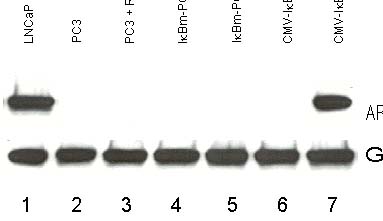
Background - (Photograph) - How can you show that three lanes are the same data? |
||
|
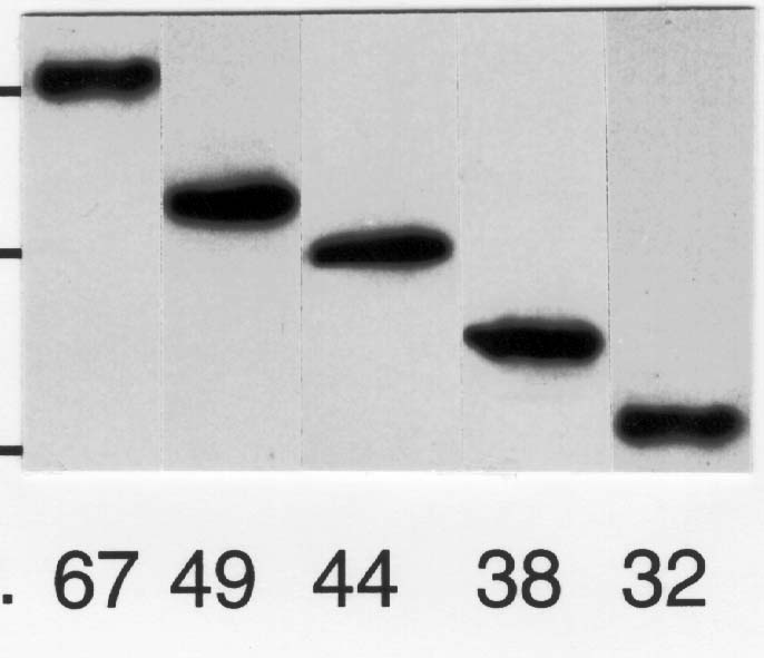
Band Detail - (Photograph) - What evidence proves the 67 kDa band is the same data as the 32 kDa band? |
||
|
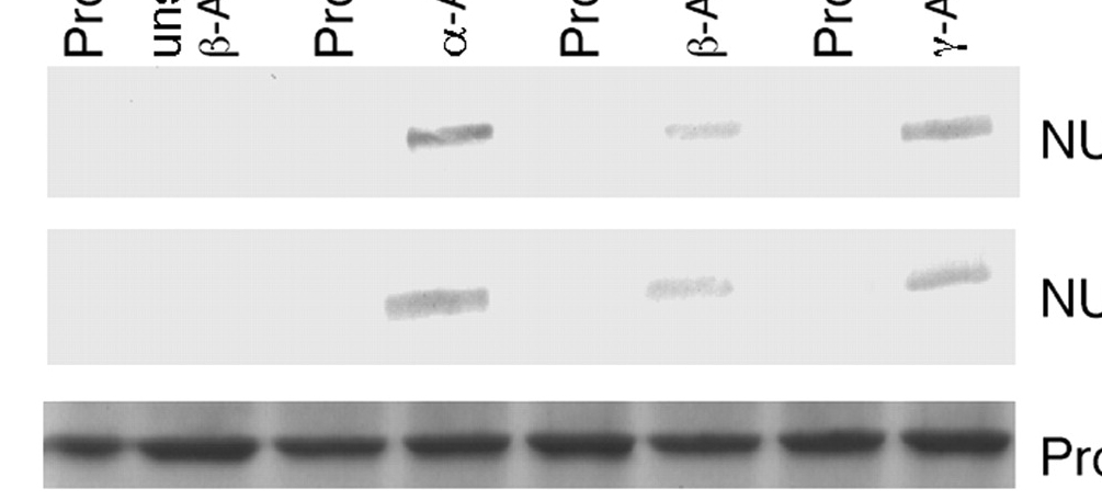
Background - Western - Which data have been fabricated - and how? - Hint, look at what's missing. |
||
|
Band Detail - JBC 2B - Do some bands look strange? - Are some the same? |
||
|
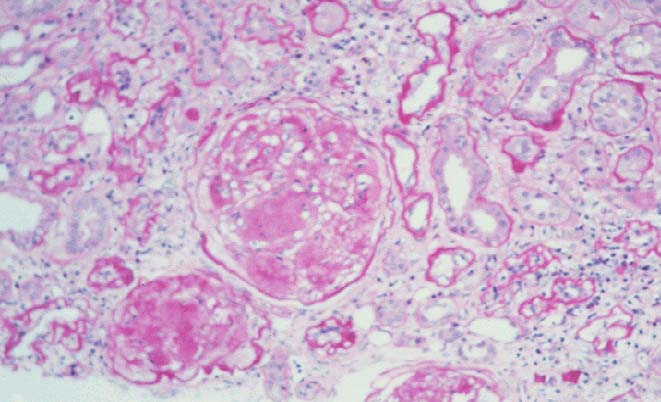
Dark Areas -Grant Figure - Can you see what's really there? |
||
|
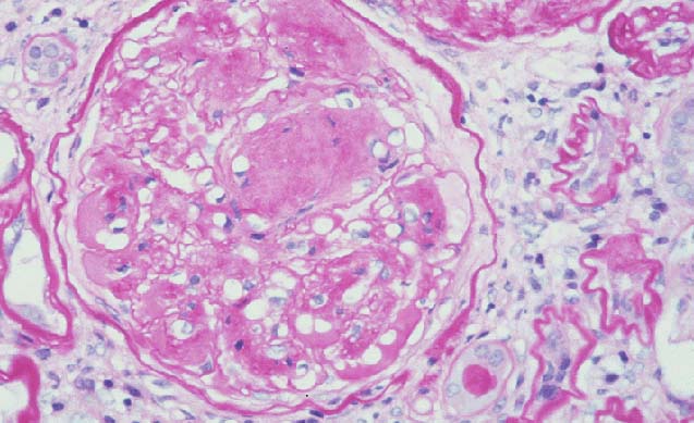
Overlap Fig A vs Fig B - Are these images from the same section? |
Fig A |
Fig B |
|
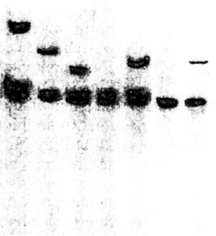
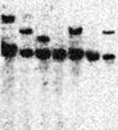
PowerPoint Slide vs "Data" - How are these "Southern blots" the same? - How do they differ? |
Slide |
"Data" |
|
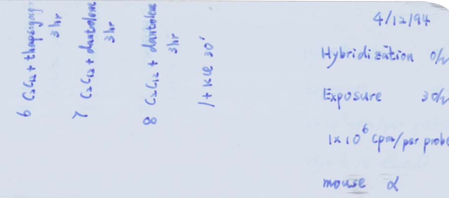
Erasures - Film - What evidence indicates that this "mouse" had feathers? - Hint, first separating by hue will improve quality of the enhanced results. |
||
Forensic-Test Patterns
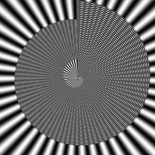
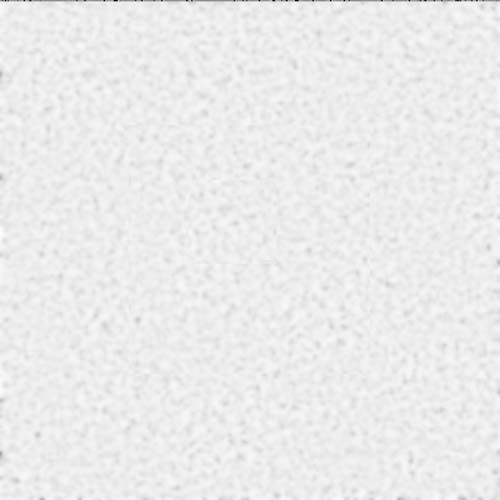
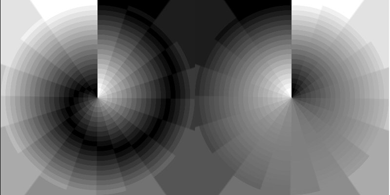
Test images with defined features are of use when trying a new processing routine, to explore whether the absence or presence of a feature (i.e., a false negative or a false positive) is not a possible artifact introduced by the routine, by the printer, or by photocopying.
The following are some trial versions.
| Description | Images | |
|---|---|---|
| Radial Resolution & Aliasing Image | ||
|
Weak Background: Duplicated & Copied Regions with Edges - Can you find the copied regions? |
||
|
Areas of Strong & Weak Contrast |
||


